Screening of halotolerant microfungi isolated from hypersaline soils of Algerian Sahara for production of hydrolytic enzymes

Submitted: October 4, 2021
Accepted: December 19, 2021
Published: December 27, 2021
Abstract Views: 2622
PDF: 635
Publisher's note
All claims expressed in this article are solely those of the authors and do not necessarily represent those of their affiliated organizations, or those of the publisher, the editors and the reviewers. Any product that may be evaluated in this article or claim that may be made by its manufacturer is not guaranteed or endorsed by the publisher.

Authors

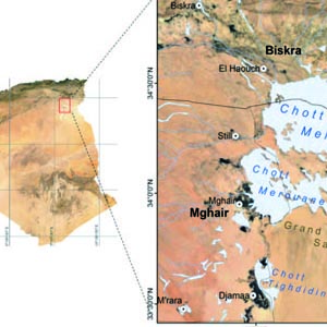
The Algerian Sahara contains numerous hypersaline ecosystems including salt lakes in which the fungal diversity has not been characterized. The abundance and diversity of soil microofungi in three salt lakes in south-eastern Algeria was investigated together with their profiles of hydrolytic enzyme. Fungal population size and relative abundance were determined in about 75 soil samples by plate count. From 69 fungal isolates, 46.38% were Aspergillus, 20.29% were Penicillium, and 11.59% belonged to Cladosporium genus. The 69 isolates have been studied at different constant temperatures and salinities. All fungal isolates are halotolerant or halophiles with the ability to grow at 50°C. The screening for extracellular halophilic enzymes at 40°C showed that 69.57% of the isolates were able to produce at least two types of the screened enzymes. Protease was the most abundant enzyme detected in 60.87% of the total isolates. The results obtained of all the growth tests indicate the adaptability of fungal isolates tested to the extreme conditions and their possible utilisation as producers of halophilic-active hydrolytic enzymes.

Dimensions

Altmetric

PlumX Metrics

Downloads

Download data is not yet available.

Citations

Oren A. Molecular ecology of extremely halophilic archaea and bacteria. FEMS Microbiol Ecol 2002;39:1-7. DOI: https://doi.org/10.1111/j.1574-6941.2002.tb00900.x
Demnati F, Samraoui B, Allache, F et al. A literature review of Algerian salt lakes: values, threats and implications. Environ Earth Sci 2017;76:127-42. DOI: https://doi.org/10.1007/s12665-017-6443-x
Mahowald NM, Bryant RG, Corral J, Steinberger L. Ephemeral lakes and desert dust sources. Geophys Res Let 2003;30:46-9. DOI: https://doi.org/10.1029/2002GL016041
Ventosa A, Nieto JJ. Biotechnological applications and potentialities of halophilic microorganisms. World J Microbiol Biotechno 1995;11:85-94. DOI: https://doi.org/10.1007/BF00339138
Anitori RP. Extremophiles: Microbiology and biotechnology. Caister Academic Press, Norfolk, UK, 2012.
Gunde-Cimerman N, Zalar P. Extremely halotolerant and halophilic fungi inhabit brine in solar salterns around the globe. Food Technol Biotechnol 2014;52:170-9.
Grishkan I, Nevo E, Wasser SP. Soil micromycete diversity in the hypersaline Dead Sea coastal area, Israel. Mycol Prog 2003;2:19-28. DOI: https://doi.org/10.1007/s11557-006-0040-9
Gunde-Cimerman N, Ramos J, Plemenitaš A. Halotolerant and halophilic fungi. Mycol Res 2009;113:1231–41. DOI: https://doi.org/10.1016/j.mycres.2009.09.002
Plemenitaš A, Vaupotič T, Lenassi M et al. Adaptation of extremely halotolerant black yeast Hortaea werneckii to increased osmolarity: a molecular perspective at a glance. Stud Mycol 2008;61:67-75. DOI: https://doi.org/10.3114/sim.2008.61.06
Zhang WW, Wang C,Xue R, Wang LJ. Effects of salinity on the soil microbial community and soil fertility. J Integr Agric 2019;18:1360-8. DOI: https://doi.org/10.1016/S2095-3119(18)62077-5
Van den Brink J, De Vries R.P. Fungal enzyme sets for plant polysaccharide degradation. Appl. Microbiol Biotechnol 2011;91:1477-92. DOI: https://doi.org/10.1007/s00253-011-3473-2
Cantrell SA, Casillas-Martinez L, Marirosa M. Characterization of fungi from hypersaline environments of solar salterns using morphological and molecular techniques. Mycol Res 2006;110:962-70. DOI: https://doi.org/10.1016/j.mycres.2006.06.005
Jaouani A, Neifar M, Prigione V, et al. Diversity and enzymatic profiling of halotolerant micromycetes from sebkha El Melah, a Saharan salt flat in southern Tunisia. Biomed Res Int 2014;2014:439197. DOI: https://doi.org/10.1155/2014/439197
Chamekh R, Deniel F, Donot C, et al. Isolation, identification and enzymatic activity of halotolerant and halophilic fungi from the great Sebkha of Oran in North western of Algeria. Mycobiology 2019;47:230-41. DOI: https://doi.org/10.1080/12298093.2019.1623979
Dendouga W, Boureghda H, Belhamra M. Edaphic factors affecting distribution of soil fungi in three Chotts located in Algerian desert. Courrier du Savoir 2015;19:147-52.
Davet P, Rouxel F. Détection et isolement des champignons du sol. INRA, Paris, 1997.
Domsch KH, Gams W, Anderson TH. Compendium of soil fungi. Academic Press London, UK; 1980.
Botton B, Breton A, Fevre M et al. Moisissures utiles et nuisibles importance industrielle. Masson, Paris; 1990.
Watanabe T. Pictorial atlas of soil and seed fungi: morphologies of cultured fungi and key to species. CRC Press, Boca Raton, USA. 2002. DOI: https://doi.org/10.1201/9781420040821
White TJ, Bruns T, Lee S, Taylor J. Amplification and direct sequencing of fungal ribosomal RNA genes for phylogenetics. In PCR Protocols: A guide to methods and applications, Innis MA, Gelfand DH, Sninsky JJ, White TJ (eds). Academic Press, San Diego, USA, 1990; pp. 315-322. DOI: https://doi.org/10.1016/B978-0-12-372180-8.50042-1
Phillips MW, Gordon GLR. Growth characteristics on cellobiose of three different anaerobic fungi isolated from the ovine rumen. Appl Environ Microbiol 1989;55:1695-1702. DOI: https://doi.org/10.1128/aem.55.7.1695-1702.1989
Hankin L, Anagnostakis SL. The use of solid media for detection of enzyme production by fungi. Mycologia 1975;67:597-607. DOI: https://doi.org/10.1080/00275514.1975.12019782
Yan N, Marschner P, Cao W, et al. Influence of salinity and water content on soil microorganisms. ISWCR 2015;3:316-23. DOI: https://doi.org/10.1016/j.iswcr.2015.11.003
Butinar L, Sonjak S, Zalar P, et al. Melanized halophilic fungi are eukaryotic members of microbial communities in hypersaline waters of solar salterns. Bot Mar 2005;48:73-9. DOI: https://doi.org/10.1515/BOT.2005.007
Molitoris HP, Buchalo AS, Kurchenko I, et al. Physiological diversity of the first filamentous fungi isolated from the hypersaline Dead Sea. Fungal Divers 2000;5:55-70.
Grishkan I, Nevo E. Spatiotemporal distribution of soil microfungi in the Makhtesh Roman area, central Negev desert. Israel Fungal Ecol 2010;3:326-37. DOI: https://doi.org/10.1016/j.funeco.2010.01.003
Kushner DJ. Microbial life in extreme environments. Academic press, London, UK; 1978.
Evans S, Hansen RW, Schneegurt MA. Isolation and characterization of halotolerant soil fungi from the great salt plains of Oklahoma. Cryptogam Mycol 2013;34:329-41. DOI: https://doi.org/10.7872/crym.v34.iss4.2013.329
Cooney DG, Emerson R. Thermophilic Fungi. W. H. Freeman and company, San Francisco; 1964.
Maheshwari R, Bharadwaj G, Bhat MK. Thermophilic fungi: their physiology and enzymes. Microbiol Mol Biol Rev 2000;64:461-88. DOI: https://doi.org/10.1128/MMBR.64.3.461-488.2000
Selbmann L, de Hoog GS, Mazzaglia A et al. Fungi at the edge of life: cryptoendolithic black fungi from Antarctic desert. Stud Mycol 2005;5:1-32.
Méjanelle L, Lòpez JF, Gunde-Cimerman N, Grimalt JO. Ergosterol biosynthesis in novel melanized fungi from hypersaline environments. J Lipid Res 2001;42:352-60. DOI: https://doi.org/10.1016/S0022-2275(20)31658-8
Plemenitaš A, Gunde-Cimerman N. Cellular responses in the halophilic black yeast Hortaea werneckii to high environmental salinity. In Adaptation to life at high salt concentrations in archaea, bacteria, and eukarya; Gunde-Cimerman N, Oren A, Plemenitaš A (eds.). Springer, Netherlands, 2005; pp. 453-470. DOI: https://doi.org/10.1007/1-4020-3633-7_29
Grishkan I. Ecological stress: Melanization as a response in fungi to radiation. In Horikoshi K, Antranikian G, Bull A, et al. (eds). Extremophiles Handbook. Springer, Tokyo, 2011; pp. 1137-1145. DOI: https://doi.org/10.1007/978-4-431-53898-1_54
Turk M, Méjanelle L, Sentjurc M et al. Salt-induced changes in lipid composition and membrane fluidity of halophilic yeast-like melanized fungi. Extremophiles 2004;8:53-61. DOI: https://doi.org/10.1007/s00792-003-0360-5
De Vries RP, Visser J. Aspergillus enzymes involved in degradation of plant cell wall polysaccharides. Microbiol Mol Biol Rev 2001;65:497-522. DOI: https://doi.org/10.1128/MMBR.65.4.497-522.2001
Tsang CC, Tang YMJ, Lau SKP, Woo PCY. Taxonomy and evolution of Aspergillus, Penicillium and Talaromyces in the omics era-Past, present and future. Comput Struct Biotechnol J 2018;16:197-210. DOI: https://doi.org/10.1016/j.csbj.2018.05.003
Meijer M, Houbraken JAMP, Dalhuijsen S et al. Growth and hydrolase profiles can be used as characteristics to distinguish Aspergillus niger and other black aspergilli. Stud Mycol 2011;69:19-30. DOI: https://doi.org/10.3114/sim.2011.69.02
Abdel-Raheem A, Shearer CA. Extracellular enzyme production by freshwater ascomycetes. Fungal Divers 2002;11:1-19.

How to Cite

Dendouga, W., & Belhamra, M. . (2021). Screening of halotolerant microfungi isolated from hypersaline soils of Algerian Sahara for production of hydrolytic enzymes. Journal of Biological Research - Bollettino Della Società Italiana Di Biologia Sperimentale, 95(1). https://doi.org/10.4081/jbr.2022.10167