Contrast-enhanced encephalopathy and massive cerebral edema after endovascular coiling of cerebral aneurysm. A case report


Published: 22 March 2021
Abstract Views: 622
PDF: 288
HTML: 2
Publisher's note
All claims expressed in this article are solely those of the authors and do not necessarily represent those of their affiliated organizations, or those of the publisher, the editors and the reviewers. Any product that may be evaluated in this article or claim that may be made by its manufacturer is not guaranteed or endorsed by the publisher.

Authors

  • Zhou Jia-hao Department of Neurosurgery, The Affiliated Hospital of Jiangsu University, Zhenjiang, Jiangsu Province, China.
  • Seidu A. Richard Department of Neurosurgery, The Affiliated Hospital of Jiangsu University, Zhenjiang, Jiangsu Province, P.R. China; Department of Medicine, Princefield University, Ho, Ghana.
  • Deng Yin-sheng Department of Neurosurgery, The Affiliated Hospital of Jiangsu University, Zhenjiang, Jiangsu Province, China.
  • Jiang Ming Department of Neurosurgery, The Affiliated Hospital of Jiangsu University, Zhenjiang, Jiangsu Province, China.
  • Zhuang Yan Department of Neurosurgery, The Affiliated Hospital of Jiangsu University, Zhenjiang, Jiangsu Province, China.

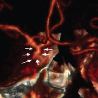
Contrast-induced encephalopathy (CIEP) is a rare complication after endovascular therapy. The etiology of CIEP is still a matter of debate. We present a rare occurrence of CIEP in a known hypertensive and type 2 diabetic patient after endovascular coiling of cerebral aneurysm with oculomotor nerve palsy. A 68-year old female presented with seven days history of headache and left ptosis or blepharoptosis with mild mydriasis. The headaches were localized mainly at the left side of the nose, orbit, and upper forehead while the left ptosis was associated with blurred vision. Computed tomography angiography revealed an aneurysm in between the C4 segment of the left internal carotid artery (ICA) and the bifurcation of the left posterior communicating artery. Digital subtraction angiography further confirmed the aneurysm. We used the transarterial approach to assess the aneurysm and subsequent coiling. Iohexol (Omnipaque) contrast agent was used during the endovascular procedure. The patient’s condition deteriorated into acute confusion state with cardinal symptomology of CIEP immediately after the operation. Computed tomography scan revealed cortical contrast enhancement in the vascular territory of the ICA as well as edema. Her symptomatology resolved 48 hours after treated with anticonvulsants, intracranial pressure reduction and hydration. Chronic hypertension as well as type 2 diabetics may be critical predisposing factors to CIEP. CIEP should be suspected in patients presenting with acute confusion state after endovascular therapy. Massive edema with ischemic brain changes in white matter of the brain before endovascular procedure should rise suspicion of CIEP.


Ikemura A, Ishibashi T, Otani K, et al. Delayed leukoencephalopathy: a rare complication after coiling of cerebral aneurysms. Am J Neuroradiol 2020;41:286-92. DOI: https://doi.org/10.3174/ajnr.A6386

Nagamine Y, Hayashi T, Kakehi Y, et al. Contrast-induced encephalopathy after coil embolization of an unruptured internal carotid artery aneurysm. Intern Med 2014;53:2133-8. DOI: https://doi.org/10.2169/internalmedicine.53.2380

Bender M, Bogdan G, Radančević D, Pejanović-Škobić N. Contrast-induced encephalopathy following cerebral angiography in a hemodialysis patient. Case Rep Neurol Med 2020;2020 [Epub ahead of print]. DOI: https://doi.org/10.1155/2020/3985231

Niimi Y, Kupersmith M, Ahmad S, et al. Cortical blindness, transient and otherwise, associated with detachable coil embolization of intracranial aneurysms. Am J Neuroradiol 2008;29:603-7. DOI: https://doi.org/10.3174/ajnr.A0858

Liao M-T, Lin T-T, Lin L-Y, et al. Contrast-induced encephalopathy after percutaneous coronary intervention. Acta Cardiol Sinica 2013;29:277.

Onishi S, Sakamoto S, Okazaki T, Kurisu K. Iodinated contrast encephalopathy after coil embolization of unruptured aneurysms. Asian J Neurosurg 2018;13:858.

Potsi S, Chourmouzi D, Moumtzouoglou A, et al. Transient contrast encephalopathy after carotid angiography mimicking diffuse subarachnoid haemorrhage. Neurol Sci 2012;33:445-8. DOI: https://doi.org/10.1007/s10072-011-0765-3

Muruve D, Steinman T. Contrast-induced encephalopathy and seizures in a patient with chronic renal insufficiency. Clin Nephrol 1996;45:406-9.

Haley Jr EC. Encephalopathy following arteriography: a possible toxic effect of contrast agents. Ann Neurol 1984;15:100-2. DOI: https://doi.org/10.1002/ana.410150118

Frantz W. Cortical blindness following coronary angiography in a patient with LIMA bypass graft and end stage renal failure. Proc Eur 2006;5:21-4.

Baguma M, Younan N, London F, et al. Contrast-associated transient cortical blindness: three cases with MRI and electrophysiology findings. Acta Neurol Belgica 2017;117:195-9. DOI: https://doi.org/10.1007/s13760-016-0696-0

Thomson KR, Varma DK. Safe use of radiographic contrast media. Aust Prescr 2010;33:35-7.

Francisco E, Seixas F, Mota A, et al. Contrast-induced encephalopathy after endovascular lower limb revascularization: a rare cause of blindness. J Anesth Clin Res 2019;10:2.

Haider AS, Gottlich C, Sumdani H, et al. Acute oculomotor nerve palsy caused by compression from an aberrant posterior communicating artery. Cureus 2019;11:e3920. DOI: https://doi.org/10.7759/cureus.3920

Esmer AF, Sen T, Comert A, et al. The neurovascular relationships of the oculomotor nerve. Clin Anat 2011;24:583-9. DOI: https://doi.org/10.1002/ca.21111

Joshi S, Tee WWH, Franconi C, Prentice D. Transient oculomotor nerve palsy due to non-aneurysmal neurovascular compression. J Clin Neurosci 2017;45:136-7. DOI: https://doi.org/10.1016/j.jocn.2017.07.006

Fang C, Leavitt JA, Hodge DO, et al. Incidence and etiologies of acquired third nerve palsy using a population-based method. JAMA Ophthalmol 2017;135:23-8. DOI: https://doi.org/10.1001/jamaophthalmol.2016.4456

Hacein-Bey L, Provenzale JM. Current imaging assessment and treatment of intracranial aneurysms. Am J Roentgenol 2011;196:32-44. DOI: https://doi.org/10.2214/AJR.10.5329

Orrù E, Roccatagliata L, Cester G, et al. Complications of endovascular treatment of cerebral aneurysms. Eur J Radiol 2013;82:1653-8. DOI: https://doi.org/10.1016/j.ejrad.2012.12.011

Suthiphosuwan S, Hsu C-T, Bharatha A. HARMless: transient cortical and sulcal hyperintensity on gadolinium-enhanced FLAIR after elective endovascular coiling of intracranial aneurysms. Am J Neuroradiol 2018;39:720-6. DOI: https://doi.org/10.3174/ajnr.A5561

Bartynski W. Posterior reversible encephalopathy syndrome, part 1: fundamental imaging and clinical features. Am J Neuroradiol 2008;29:1036-42. DOI: https://doi.org/10.3174/ajnr.A0928

Bartynski W. Posterior reversible encephalopathy syndrome, part 2: controversies surrounding pathophysiology of vasogenic edema. Am J Neuroradiol 2008;29:1043-9. DOI: https://doi.org/10.3174/ajnr.A0929

Leong S, Fanning N. Persistent neurological deficit from iodinated contrast encephalopathy following intracranial aneurysm coiling: a case report and review of the literature. Interv Neuroradiol 2012;18:33-41. DOI: https://doi.org/10.1177/159101991201800105

Sawaya RA, Hammoud R, Arnaout S, Alam S. Contrast-induced encephalopathy following coronary angioplasty with iohexol. Southern Med J 2007;100:1054-5. DOI: https://doi.org/10.1097/SMJ.0b013e3181540086

Uchiyama Y, Abe T, Hirohata M, et al. Blood brain-barrier disruption of nonionic iodinated contrast medium following coil embolization of a ruptured intracerebral aneurysm. Am J Neuroradiol 2004;25:1783-6.

Junck L, Marshall W. Fatal brain edema after contrast-agent overdose. Am J Neuroradiol 1986;7:522-5.

Zhao W, Zhang J, Song Y, et al. Irreversible fatal contrast-induced encephalopathy: a case report. BMC Neurol 2019;19:46. DOI: https://doi.org/10.1186/s12883-019-1279-5

Mentzel H-J, Blume J, Malich A, et al. Cortical blindness after contrast-enhanced CT: complication in a patient with diabetes insipidus. Am J Neuroradiol 2003;24:1114-6.

Matsubara N, Izumi T, Miyachi S, et al. Contrast-induced encephalopathy following embolization of intracranial aneurysms in hemodialysis patients. Neurol Med-Chir 2017:oa.2017-0132. DOI: https://doi.org/10.2176/nmc.oa.2017-0132

Shinoda J, Ajimi Y, Yamada M, Onozuka S. Cortical blindness during coil embolization of an unruptured intracranial aneurysm. Neurol Med-Chir 2004;44:416-9. DOI: https://doi.org/10.2176/nmc.44.416

Merchut MP, Richie B. Transient visuospatial disorder from angiographic contrast. Archiv Neurol 2002;59:851-4.

Yu J, George Dangas M. New insights into the risk factors of contrast-induced encephalopathy. J Endovasc Ther 2011;18:545. DOI: https://doi.org/10.1583/11-3476C.1

Jia-hao, Z. ., Richard, S. A., Yin-sheng, D. ., Ming, J., & Yan, Z. (2021). Contrast-enhanced encephalopathy and massive cerebral edema after endovascular coiling of cerebral aneurysm. A case report. Veins and Lymphatics, 10(1). https://doi.org/10.4081/vl.2021.9767

Downloads

Download data is not yet available.

Citations