Evaluation of paraclinical methods in diagnosis of Foreign Body and its complications compared to operating room findings in patients with oral, pharyngeal and esophageal symptoms in Amir A’lam Hospital


Submitted: 2 December 2019
Accepted: 28 February 2020
Published: 8 May 2020
Abstract Views: 1407
PDF: 276
Publisher's note
All claims expressed in this article are solely those of the authors and do not necessarily represent those of their affiliated organizations, or those of the publisher, the editors and the reviewers. Any product that may be evaluated in this article or claim that may be made by its manufacturer is not guaranteed or endorsed by the publisher.

Authors

  • Reza Erfanian Otorhinolaryngology Research Center, Tehran University of Medical Sciences, Tehran, Iran, Islamic Republic of.
  • Ardavan Tajadini Otorhinolaryngology Research Center, Tehran University of Medical Sciences, Tehran, Iran, Islamic Republic of.
  • Saeed Sohrabpour Otorhinolaryngology Research Center, Tehran University of Medical Sciences, Tehran, Iran, Islamic Republic of.
  • Keyvan Aghazadeh Otorhinolaryngology Research Center, Tehran University of Medical Sciences, Tehran, Iran, Iran, Islamic Republic of.
  • Abdolhossein Mehdinezhad Otorhinolaryngology Research Center, Tehran University of Medical Sciences, Tehran, Iran, Islamic Republic of.
  • Ebrahim Karimi Otorhinolaryngology Research Center, Tehran University of Medical Sciences, Tehran, Iran, Islamic Republic of.

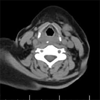
Foreign Body (FB) swallowing is one of the most common medical emergencies in the world. This injury can cause mortality and morbidity in all age groups. This study aimed to evaluate the paraclinical methods in diagnosis of foreign body and its complications compared to operating room findings in patients with oral, pharyngeal and esophageal symptoms. This was a retrospective study. All patients referred to the ENT (Ear, Nose, Throat) department of Amir A’lam hospital with complaints of foreign body swallowing admitted in the operating room were studied. The patients’ information including demographic information, type of swallowed body, paraclinical information and operating room findings were extracted from the patients’ records. This study was performed on 219 patients admitted to ENT clinic. The mean age of the patients was 45.10±20.61 years. The highest and the lowest frequencies for foreign body type were chicken bone in 61 cases (28.31%) and disk battery in 1 case (0.5%). The most and the least frequency of complaints after foreign body type were dysphagia in 129 cases (58.9%) and neck tenderness in 7 cases (3.19%). Computerized Tomography (CT) scan findings and operative findings were almost same except in two cases. Kappa agreement for lateral neck and observation in the operating room was 50.5% (P <0.001) and WBC count was significantly higher in patients with complications (P=0.010).


Guirgis M, Nguyen R, Pokorny C. Accidental ingestion of plastic from takeaway containers--food for thought. Med J Aust. 2011 Mar; 194(5): 245-46. 2. Okhakhu AL, Ogisi FO. An unusual foreign body in human oesophagus – case report. Benin Journsl of Postgraduate Medicine. 2007 Dec; 9(1): 41-43.

Baraka A, Bikhazi G. Oesophageal foreign bodies. Br Med J. 1975 Mar; 1(5957): 561-63. DOI: https://doi.org/10.1136/bmj.1.5957.561

Baral BK, Joshi RR, Bhattarai BK, Sewal RB. Removal of coin from upper esophageal tract in children with Magill’s forceps under propofol sedation. Nepal Med Coll J. 2010 Mar;12(1):38-41. DOI: https://doi.org/10.1007/s10039-009-1543-9

Boedeker BH, Bernhagen MA, Miller DJ, Doyle DJ. Comparison of the Magill forceps and the Boedeker (curved) intubation forceps for removal of a foreign body in a Manikin. Journal of clinical anesthesia. 2012 Feb 1;24(1):25-7. DOI: https://doi.org/10.1016/j.jclinane.2011.04.013

Akenroye MI, Osukoya AT. Uncommon, undeclared oesophageal foreign bodies. Niger J Clin Pract. 2012 Apr-Jun; 15(2): 244-46. doi: 10.4103/1119-3077.97339. DOI: https://doi.org/10.4103/1119-3077.97339

Vyas K, Sawant P, Rathi P, Das HS, Borse N. Foreign bodies in gut. J Assoc Physicians India. 2000 Apr; 48(4): 394- 96.

Shivakumar AM, Naik AS, Parashonth KB, Hongal GF, Chaturvedy G. Foreign bodies in upper digestive tract. Indian J Otolaryngol Head Neck Surg. 2006 Jan; 58(1): 63–68. doi: 9.1007/BF02907744 7.

Pak MW, Lee WC,Fung HK, van Hassect CA. A prospective study of foreign- body ingestion in 311 children. Int J Pediatr Otorhinolaryngol. 2001 Apr; 58(1): 37-45. https://doi.org/10.1016/S0165-5876(00) 00464-X.

Vargas EJ, Mody AP, Kim TY, Denmark TK, Moynihan JA, Barcega BB, Khan A, Clark RT, Brown L. The removal of coins from the upper esophageal tract of children by emergency physicians: a pilot study. Canadian Journal of Emergency Medicine. 2004 Nov;6(6):434-40. DOI: https://doi.org/10.1017/S1481803500009453

Higgins GL, Burton JH, Carter WP, Floor AE. Comparison of Extraction Devices for the Removal of Supraglottic Foreign Bodies. Prehospital Emergency Care. 2003 Jan 1;7(3):316-21. DOI: https://doi.org/10.1080/10903120390936491

Nouruzi-Sedeh P, Schumann M, Groeben H. Laryngoscopy via Macintosh Bladeversus GlideScope. Anesthesiology 2009;110:32–7. DOI: https://doi.org/10.1097/ALN.0b013e318190b6a7

Xue FS, Yuan YJ, Liao X, Xiong J, Wang Q. Is Glidescope® videolaryngoscope more effective than Macintosh laryngoscope for emergent intubation during chest compression?. Resuscitation. 2011 Jul 1;82(7):956. DOI: https://doi.org/10.1016/j.resuscitation.2011.02.049

Damghani M, Halavati N, Motamedi N. Foreign body in the upper airway and oesophagus: a seven years study from Iran. J Pak Med Assoc. 2011 Sep;61(9 (:859-62.

Cheng W, Tam PK. Foreign-body ingestion in children: experience with 1,265 cases. J Pediatr Surg. 1999 Oct; 34(10): 1472-76. DOI: https://doi.org/10.1016/S0022-3468(99)90106-9

Flint PW, Haughey BH, Niparko JK, Richardson MA, Lund VJ, Robbins KT, Lesperance MM, Thomas JR. Cummings Otolaryngology-Head and Neck Surgery E-Book: Head and Neck Surgery, 3-Volume Set. Elsevier Health Sciences; 2010 Mar 9.

Kliegmassn RM, Stanton B, Geme JS, Schor NF, Behrman RE. Nelson textbook of pediatrics.20 ed. Elsevier 2015; Vol 2.p 2039.

Brunicardi F, Andersen D, Billiar T, Dunn D, Hunter J, Matthews J, Pollock R. Schwartz's principles of surgery, 10e. McGraw-hill; 2014.

Sharafi A, Taziki MH, Razaei S. Prevalence of esophageal foreign bodies and its complications in Gorgan, northern Iran (2004-14). Journal of Gorgan University of Medical Sciences. 2017 Sep 1;19 (3).

Karimaneh A, Najafi M. [Esophageal foreign bodies]. Pajhohesh dar Pezeshki. 2004; 28(1): 49-52. [Persian]

Pirzadeh A, Mahdavi A, Charkhtab J. [A survey on the esophagus foreign bodies among patients in Fatemy hospital, Ardabil, 1998-2001]. J Ardabil Univ Med Sci. 2003; 3(1): 13-17. [Persian]

Pourrashidi R, Sharifkashani S, Sharifian H, Mazaher H, Salamati P, Ghorbani yekta B. Comparison of ultrasonography with radiography for the detection of cervical esophageal foreign body. Tehran Univ Med J. 2013; 71 (4) :224-229.

Sung SH, Jeon SW, Son HS, Kim SK, Jung MK, Cho CM, Tak WY, Kweon YO. Factors predictive of risk for complications in patients with oesophageal foreign bodies. Digestive and Liver Disease. 2011 Aug 1;43(8):632-5. DOI: https://doi.org/10.1016/j.dld.2011.02.018

Santucci CA, Purcell TB, Mejia C. Leukocytosis as a predictor of severe injury in blunt trauma. Western Journal of Emergency Medicine. 2008 May; 9(2):81.

Erfanian, R., Tajadini, A., Sohrabpour, S., Aghazadeh, K., Mehdinezhad, A., & Karimi, E. (2020). Evaluation of paraclinical methods in diagnosis of Foreign Body and its complications compared to operating room findings in patients with oral, pharyngeal and esophageal symptoms in Amir A’lam Hospital. Emergency Care Journal, 16(1). https://doi.org/10.4081/ecj.2020.8726

Downloads

Download data is not yet available.

Citations