Accuracy of bedside sonographic measurement of optic nerve sheath diameter for intracranial hypertension diagnosis in the emergency department
Submitted: 23 March 2023
Accepted: 8 May 2023
Published: 23 June 2023
Abstract Views: 1044
PDF: 310
Publisher's note
All claims expressed in this article are solely those of the authors and do not necessarily represent those of their affiliated organizations, or those of the publisher, the editors and the reviewers. Any product that may be evaluated in this article or claim that may be made by its manufacturer is not guaranteed or endorsed by the publisher.

Authors

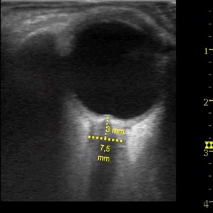
Ultrasound measurement of the optic nerve sheath diameter (US ONSD) has been proposed as a method to diagnose elevated intracranial pressure (EICP), but the optimal threshold is unclear. The aim of this study was to assess the accuracy of US ONSD, as compared to head computed tomography (CT), in detecting EICP of both traumatic and non-traumatic origin. We conducted a prospective, cross-sectional, multicenter study. Patients presenting to the emergency department with a suspect of traumatic or non-traumatic brain injury, referred for an urgent head CT, underwent US ONSD measurement. A US ONSD ≥5.5 mm was considered positive. Sensitivity, specificity, positive and negative predictive values, and positive and negative likelihood ratios were calculated for three ONSD cut-offs: 5.5 (primary outcome), 5.0, and 6.0 mm. A receiver operating characteristic (ROC) curve was also generated and the area under the ROC curve calculated. Ninetynine patients were enrolled. The CT was positive in 15% of cases and the US ONSD was positive in all of these, achieving a sensitivity of 100% [95% confidence interval (CI) 78; 100] and a negative predictive value of 100% (95% CI 79; 100). The CT was negative in 85% of cases, while the US ONSD was positive in 69% of these, achieving a specificity of 19% (95% CI 11; 29) and a positive predictive value of 18% (95% CI 11; 28). The US ONSD, with a 5.5 mm cut-off, might safely be used to rule out EICP in patients with traumatic and non-traumatic brain injury in the ED. In limited-resources contexts, a negative US ONSD could allow emergency physicians to rule out EICP in low-risk patients, deferring the head CT.

Dimensions

Altmetric

PlumX Metrics

Downloads

Download data is not yet available.

Citations

Changa AR, Czeisler BM, Lord AS. Management of Elevated Intracranial Pressure: a Review. Curr Neurol Neurosci Rep 2019;19:99. DOI: https://doi.org/10.1007/s11910-019-1010-3
Mollan SP, Spitzer D, Nicholl DJ. Raised intracranial pressure in those presenting with headache. BMJ. 2018 Oct 4;363:k3252. DOI: https://doi.org/10.1136/bmj.k3252
Citerio G, Andrews PJ. Intracranial pressure. Part two: Clinical applications and technology. Intensive Care Med 2004;30:1882-5. DOI: https://doi.org/10.1007/s00134-004-2377-3
Fried HI, Nathan BR, Rowe AS, et al. The insertion and management of external ventricular drains: an evidence-based consensus statement: a statement for healthcare professionals from the Neurocritical Care Society. Neurocrit Care 2016;24:61-81. DOI: https://doi.org/10.1007/s12028-015-0224-8
Tavakoli S, Peitz G 3rd, Ares W, et al. Complications of invasive intracranial pressure monitoring devices in neurocritical care. Neurosurg Focus 2017;43:E6. DOI: https://doi.org/10.3171/2017.8.FOCUS17450
Binz DD, Toussaint LG, Friedman JA. Hemorrhagic complications of ventriculostomy placement: a meta-analysis. Neurocrit Care 2009;10:253-6. DOI: https://doi.org/10.1007/s12028-009-9193-0
Holloway KL, Barnes T, Choi S, et al. Ventriculostomy infections:the effect of monitoring duration and catheter exchange in 584 patients. J Neurosurg 1996;85:419-24. DOI: https://doi.org/10.3171/jns.1996.85.3.0419
Raboel PH, Bartek J Jr, Andresen M, et al. Intracranial pressure monitoring: invasive versus non-invasive methods—a review. Crit Care Res Pract 2012;2012:950393. DOI: https://doi.org/10.1155/2012/950393
Eisenberg HM, Gary HE Jr, Aldrich EF, et al. Initial CT findings in 753 patients with severe head injury. A report from the NIH Traumatic Coma Data Bank. J Neurosurg 1990;73:688-98. DOI: https://doi.org/10.3171/jns.1990.73.5.0688
Tayal VS, Neulander M, Norton HJ, et al. Emergency department sonographic measurement of optic nerve sheath diameter to detect findings of increased intracranial pressure in adult head injury patients. Ann Emerg Med 2007;49:508-14. DOI: https://doi.org/10.1016/j.annemergmed.2006.06.040
Koziarz A, Sne N, Kegel F, et al. Optic nerve sheath diameter sonography for the diagnosis of increased intracranial pressure: A systematic review and meta-analysis protocol. BMJ 2017;7:e016194. DOI: https://doi.org/10.1136/bmjopen-2017-016194
Ballantyne SA, O’Neill G, Hamilton R, et al. Observer variation in the sonographic measurement of optic nerve sheath diameter in normal adults. Eur J Ultrasound 2002;15:145-9. DOI: https://doi.org/10.1016/S0929-8266(02)00036-8
Koziarz A, Sne N, Kegel F, et al.. Bedside Optic Nerve Ultrasonography for Diagnosing Increased Intracranial Pressure. A Systematic Review and Meta-analysis. Ann Intern Med 2019;171:896-905. DOI: https://doi.org/10.7326/M19-0812
Dubourg J, Javouhey E, Geeraerts T, Messerer M, Kassai B. Ultrasonography of optic nerve sheath diameter for detection of raised intracranial pressure: a systematic review and meta-analysis. Intensive Care Med 2011;37:1059-68. DOI: https://doi.org/10.1007/s00134-011-2224-2
Kimberly HH, Shah S, Marill K, Noble V. Correlation of optic nerve sheath diameter with direct measurement of intracranial pressure. Acad Emerg Med 2008;15:201-4. DOI: https://doi.org/10.1111/j.1553-2712.2007.00031.x
Rajajee V, Vanaman M, Fletcher JJ, Jacobs TL. Optic nerve ultrasound for the detection of raised intracranial pressure. Neurocrit Care 2011;15:506-15. DOI: https://doi.org/10.1007/s12028-011-9606-8
Robba C, Santori G, Czosnyka M, et al. Optic nerve sheath diameter measured sonographically as non-invasive estimator of intracranial pressure: a systematic review and meta-analysis. Intensive Care Med 2018;44:1284-94. DOI: https://doi.org/10.1007/s00134-018-5305-7
Liu D, Li Z, Zhang X, et al. Assessment of intracranial pressure with ultrasonographic retrobulbar optic nerve sheath diameter measurement. BMC Neurol 2017;17:188. DOI: https://doi.org/10.1186/s12883-017-0964-5
Lochner P, Czosnyka M, Naldi A, et al. Optic nerve sheath diameter: present and future perspectives for neurologists and critical care physicians. Neurol Sci 2019;40:2447-57. DOI: https://doi.org/10.1007/s10072-019-04015-x
Hylkema C. Optic Nerve Sheath Diameter Ultrasound and the Diagnosis of Increased Intracranial Pressure. Crit Care Nurs Clin North Am 2016;28:95-9. DOI: https://doi.org/10.1016/j.cnc.2015.10.005
Karakitsos D, Soldatos T, Gouliamos A, et al. Transorbital sonographic monitoring of optic nerve diameter in patients with severe brain injury. Transplant Proc 2006;38:3700-6. DOI: https://doi.org/10.1016/j.transproceed.2006.10.185
Ohle R, McIsaac SM, Woo MY, Perry JJ. Sonography of the optic nerve sheath diameter for detection of raised intracranial pressure compared to computed tomography: a systematic review and meta-analysis. J Ultrasound Med 2015;34:1285-94. DOI: https://doi.org/10.7863/ultra.34.7.1285
Soldatos T, Chatzimichail K, Papathanasiou M, Gouliamos A. Optic nerve sonography: a new window for the non-invasive evaluation of intracranial pressure in brain injury. Emerg Med J 2009;26:630-4. DOI: https://doi.org/10.1136/emj.2008.058453
Neri L, Storti E, Lichtenstein D. Toward an ultrasound curriculum for critical care medicine. Crit Care Med 2007;35:S290-304. DOI: https://doi.org/10.1097/01.CCM.0000260680.16213.26
Soroushmehr R, Rajajee K, Williamson C, et al. Automated optic nerve sheath diameter measurement using super-pixel analysis. Annu Int Conf IEEE Eng Med Biol Soc 2019;2019:2793-6. DOI: https://doi.org/10.1109/EMBC.2019.8856449
Kim SE, Hong EP, Kim HC, et al. Ultrasonographic optic nerve sheath diameter to detect increased intracranial pressure in adults: a meta-analysis. Acta Radiol 2019;60:221-9. DOI: https://doi.org/10.1177/0284185118776501
Nakagawa K, Smith WS. Evaluation and management of increased intracranial pressure. Continuum (Minneap Minn) 2011;17:1077-93. DOI: https://doi.org/10.1212/01.CON.0000407061.25284.28
Servadei F, Murray GD, Penny K, et al. The value of the "worst" computed tomographic scan in clinical studies of moderate and severe head injury. Neurosurgery 2000;46:70-5; discussion 75-7. DOI: https://doi.org/10.1093/neurosurgery/46.1.70
Vaiman M, Gottlieb P, Bekerman I. Quantitative relations between the eyeball, the optic nerve, and the optic canal important for intracranial pressure monitoring. Head Face Med. 2014;10:32. DOI: https://doi.org/10.1186/1746-160X-10-32

How to Cite

Busti, C., Marcosignori , M., Marchetti , F., Batori , G., Giovenali , L., Corea , F., Calabrò , G., Monti, M., & Germini , F. (2023). Accuracy of bedside sonographic measurement of optic nerve sheath diameter for intracranial hypertension diagnosis in the emergency department. Emergency Care Journal, 19(2). https://doi.org/10.4081/ecj.2023.11333