A direct comparison between five lung-US and chest-CT-scans in a patient infected by SARS-CoV-2
Submitted: 2 April 2022
Accepted: 10 August 2022
Published: 27 September 2022
Abstract Views: 572
PDF: 230
Videoclip 1: 0
Videoclip 2: 0
Videoclip 3: 0
Videoclip 4: 0
Publisher's note
All claims expressed in this article are solely those of the authors and do not necessarily represent those of their affiliated organizations, or those of the publisher, the editors and the reviewers. Any product that may be evaluated in this article or claim that may be made by its manufacturer is not guaranteed or endorsed by the publisher.

Authors

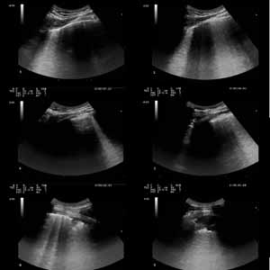
The gold standard for SARS-CoV-2 pneumonia diagnosis is chest Computed Tomography (CT), but Lung Ultrasound (LUS) is also useful in differential diagnosis and in-hospital monitoring of patients with infection by new Coronavirus 2019 disease (COVID- 19). We present a case of a young man who was infected with SARS-CoV-2 pneumoniae and underwent five steps of chest imaging, including LUS aeration scorings and chest CT scans. Each decrease or increase in LUS scoring could accurately predict CT scan changes.

Dimensions

Altmetric

PlumX Metrics

Downloads

Download data is not yet available.

Citations

Ye Z, Zhang Y, Wang Y, Huang Z, Song B. Chest CT manifestations of new coronavirus disease 2019 (COVID-19): a pictorial review. Eur Radiol 2020;30:4381-9. DOI: https://doi.org/10.1007/s00330-020-06801-0
Simpson S KF, Abbara S, Bhalla S, et al. Radiological Society of North America Expert Consensus Statement on Reporting Chest CT Findings Related to COVID-19. Endorsed by the Society of Thoracic Radiology, the American College of Radiology, and RSNA - Secondary Publication. J Thorac Imaging 2020:35:219-27. DOI: https://doi.org/10.1097/RTI.0000000000000524
Volpicelli G, Gargani L. Sonographic signs and patterns of COVID-19 pneumonia. Ultrasound J 2020:12:22. DOI: https://doi.org/10.1186/s13089-020-00171-w
Gargani L, Soliman-Aboumarie H, Volpicelli G, et al. Why, when, and how to use lung ultrasound during the COVID-19 pandemic: enthusiasm and caution. Eur Heart J Cardiovasc Imaging 2020;21:941-8. DOI: https://doi.org/10.1093/ehjci/jeaa163
Volpicelli G, Gargani L, Perlini S, et al. Lung ultrasound for the early diagnosis of COVID-19 pneumonia: an international multicenter study. Intensive Care Med 2021;47:444-54. DOI: https://doi.org/10.1007/s00134-021-06373-7
Zieleskiewicz L, Markarian T, Lopez A, et al. Comparative study of lung ultrasound and chest computed tomography scan in the assessment of severity of confirmed COVID-19 pneumonia. Intensive Care Med 2020;46:1707-13. DOI: https://doi.org/10.1007/s00134-020-06186-0
Nouvenne A, Zani MD, Milanese G, et al. Lung ultrasound in COVID-19 pneumonia: correlations with chest CT on hospital admission. Respiration 2020;99:617-24. DOI: https://doi.org/10.1159/000509223
Bouhemad B, Mongodi S, Via G, Rouquette I. Ultrasound for "lung monitoring" of ventilated patients. Anesthesiology 2015;122:437-47. DOI: https://doi.org/10.1097/ALN.0000000000000558
Chung M, Bernheim A, Mei X, et al. CT imaging features of 2019 novel coronavirus (2019-nCoV). Radiology 2020;295:202-7. DOI: https://doi.org/10.1148/radiol.2020200230
Falcone M, Tiseo G, Barbieri G, et al. Role of low-molecular-weight heparin in hospitalized patients with severe acute respiratory syndrome coronavirus 2 pneumonia: a prospective observational study. Open Forum Infect Dis 2020;7:ofaa563. DOI: https://doi.org/10.1093/ofid/ofaa563
Soldati G SA, Inchingolo R, Buonsenso D, et al. Proposal for international standardization of the use of lung ultrasound for patients with COVID-19: a simple, quantitative, reproducible method. J Ultrasound Med 2020;39:1413-9. DOI: https://doi.org/10.1002/jum.15285
Boccatonda A, Grignaschi A, Lanotte AMG, et al. Role of lung ultrasound in the management of patients with suspected SARS-CoV-2 infection in the emergency department. J Clin Med 2022;11:2067 DOI: https://doi.org/10.3390/jcm11082067
Montanari M, De Ciantis P, Boccatonda A, et al. Lung ultrasound monitoring of CPAP effectiveness on SARS-CoV-2 pneumonia: A case report. Emer Care J 2020;16:9333. DOI: https://doi.org/10.4081/ecj.2020.9333
Boccatonda A, Cocco G, Ianniello E, et al. One year of SARS-CoV-2 and lung ultrasound: what has been learned and future perspectives. J Ultrasound 2021;24:115-23. DOI: https://doi.org/10.1007/s40477-021-00575-x
Boccatonda A, Cocco G, Sofia S, et al. A new type of outpatient: lung ultrasound after covid-19 infection. J Ultrasound Med 2022;41:2113-14. DOI: https://doi.org/10.1002/jum.15871

How to Cite

Poli, V., Spinelli, S., Toscano, M., Vitale, F., Ranalli, A., Tonerini, M., Romei, C., De Liperi, A., & Barbieri, G. (2022). A direct comparison between five lung-US and chest-CT-scans in a patient infected by SARS-CoV-2. Emergency Care Journal, 18(3). https://doi.org/10.4081/ecj.2022.10492

Similar Articles

You may also start an advanced similarity search for this article.