COVID-19 and cutaneous manifestations: Two cases and a review of the literature

Submitted: 18 March 2022
Accepted: 13 April 2022
Published: 27 June 2022
Abstract Views: 934
PDF: 208
Publisher's note
All claims expressed in this article are solely those of the authors and do not necessarily represent those of their affiliated organizations, or those of the publisher, the editors and the reviewers. Any product that may be evaluated in this article or claim that may be made by its manufacturer is not guaranteed or endorsed by the publisher.

Authors

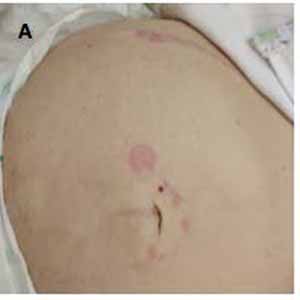
COVID-19 can affect multiple organs, including skin. A wide range of skin manifestations have been reported in literature. Six main phenotypes have been identified: i) urticarial rash, ii) confluent erythematous/maculopapular/morbilliform rash, iii) papulovesicular exanthem, iv) a chilblain-like acral pattern, v) a livedo reticularis/racemosa-like pattern, and vi) a purpuric vasculitic pattern. The pathogenetic mechanism is still not completely clear, but a role of hyperactive immune response, complement activation and microvascular injury have been postulated. The only correlation between the cutaneous phenotype and the severity of COVID-19 has been observed in the case of chilblain-like acral lesions, that is generally associated with the benign/subclinical course of COVID-19. Herein, we report two cases of SARS-CoV- 2 infection in patients who developed cutaneous manifestations that completely solved with systemic steroids and antihistamines. The first case is a female patient not vaccinated for SARS-CoV-2 with COVID-19 associated pneumonia, while the second case is a vaccinated female patient with only skin manifestations.

Dimensions

Altmetric

PlumX Metrics

Downloads

Download data is not yet available.

Citations

Molina-Ruiz AM, Santonja C, Rütten A, et al. Immunohistochemistry in the diagnosis of cutaneous viral infections--part I. Cutaneous viral infections by herpesviruses and papillomaviruses. Am J Dermatopathol 2015;37:1-14. DOI: https://doi.org/10.1097/DAD.0000000000000203
Muehlenbachs A, Bhatnagar J, Zaki SR. Tissue tropism, pathology and pathogenesis of enterovirus infection. J Pathol 2015;235:217-28. DOI: https://doi.org/10.1002/path.4438
Keighley CL, Saunderson RB, Kok J, Dwyer DE. Viral exanthems. Curr Opin Infect Dis 2015;28:139-50. DOI: https://doi.org/10.1097/QCO.0000000000000145
Su CJ, Lee CH. Viral exanthem in COVID-19, a clinical enigma with biological significance. J Eur Acad Dermatol Venereol 2020;34:e251-2. DOI: https://doi.org/10.1111/jdv.16469
Recalcati S. Cutaneous manifestations in COVID-19: a first perspective. J Eur Acad Dermatol Venereol 2020;34:e212–3. DOI: https://doi.org/10.1111/jdv.16387
Guan WJ, Ni ZY, Hu Y, et al. China medical treatment expert group for Covid-19. Clinical characteristics of Coronavirus Disease 2019 in China. N Engl J Med 2020;382:1708-20. DOI: https://doi.org/10.1056/NEJMc2005203
Hoehl S, Rabenau H, Berger A, et al. Evidence of SARS-CoV-2 infection in returning travelers from Wuhan, China. N Engl J Med 2020;382:1278-80. DOI: https://doi.org/10.1056/NEJMc2001899
Joob B, Wiwanitkit V. COVID-19 can present with a rash and be mistaken for dengue. J Am Acad Dermatol 2020;82:e177. DOI: https://doi.org/10.1016/j.jaad.2020.03.036
Kamali Aghdam M, Jafari N, Eftekhari K. Novel coronavirus in a 15-day-old neonate with clinical signs of sepsis, a case report. Infect Dis (Lond) 2020;52:427-9. DOI: https://doi.org/10.1080/23744235.2020.1747634
Uchonwanit P, Leerunyakul K, Kositkuljorn C. Cutaneous manifestations in COVID-19: Lessons learned from current evidence. J Am Acad Dermatol 2020;83:e57-e60. DOI: https://doi.org/10.1016/j.jaad.2020.04.094
De Giorgi V, Recalcati S, Jia Z, et al. Cutaneous manifestations related to coronavirus disease 2019 (COVID-19): a prospective study from China and Italy. J Am Acad Dermatol 2020;83:674–5. DOI: https://doi.org/10.1016/j.jaad.2020.05.073
Galvan Casas C, Catala A, Carretero Hernandez G, et al. Classification of the cutaneous manifestations of COVID-19: a rapid prospective nationwide consensus study in Spain with 375 cases. Br J Dermatol 2020;183:71-7. DOI: https://doi.org/10.1111/bjd.19347
Marzano AV, Cassano N, Genovese G, et al. Cutaneous manifestations in patients with COVID-19: a preliminary review of an emerging issue. Br J Dermatol 2020;183:431-42. DOI: https://doi.org/10.1111/bjd.19264
Torrelo A, Andina D, Santonja C, et al. Erythema multiformelike lesions in children and COVID-19. Pediatr Dermatol 2020;37:442-6. DOI: https://doi.org/10.1111/pde.14246
Ehsani AH, Nasimi M, Bigdelo Z. Pityriasis rosea as a cutaneous manifestation of COVID-19 infection. J Eur Acad Dermatol Venereol 2020;34:e436-7. DOI: https://doi.org/10.1111/jdv.16579
Llamas-Velasco M, Chicharro P, Rodríguez Jiménez P, et al. Comment on ‘Clinical and histological characterization of vesicular COVID-19 rashes: a prospective study in a tertiary care hospital’. Pseudoherpetic Grover disease seems to occur in patients with COVID-19 infection. Clin Exp Dermatol 2020;45:896–8. DOI: https://doi.org/10.1111/ced.14305
Cepeda-Valdes R, Carrion-Alvarez D, Trejo Castro A, et al. Cutaneous manifestations in COVID-19: familial cluster of urticarial rash. Clin Exp Dermatol 2020;45:895–6. DOI: https://doi.org/10.1111/ced.14290
Freeman EE, McMahon DE, Lipoff JB, et al. The spectrum of COVID-19-associated dermatologic manifestations: an international registry of 716 patients from 31 countries. J Am Acad Dermatol 2020;83:1118–29. DOI: https://doi.org/10.1016/j.jaad.2020.06.1016
Najafzadeh M, Shahzad F, Ghaderi N, et al. Urticaria (angioedema) and COVID-19 infection. J Eur Acad Dermatol Venereol 2020;34:e568–70. DOI: https://doi.org/10.1111/jdv.16721
Fernandez-Nieto D, Ortega-Quijano D, Jimenez-Cauhe J, et al. Clinical and histological characterization of vesicular COVID-19 rashes: a prospective study in a tertiary care hospital. Clin Exp Dermatol 2020;45:872–5. DOI: https://doi.org/10.1111/ced.14277
Amatore F, Macagno N, Mailhe M, Demarez B, Gaudy-Marqueste C, Grob JJ, Raoult D, Brouqui P, Richard MA. SARS-CoV-2 infection presenting as a febrile rash. J Eur Acad Dermatol Venereol 2020;34:e304-e306. DOI: https://doi.org/10.1111/jdv.16528
Rodríguez-Jiménez P, Chicharro P, De Argila D, et al. Urticaria-like lesions in COVID-19 patients are not really urticaria - a case with clinicopathological correlation. J Eur Acad Dermatol Venereol 2020;34:e459-60. DOI: https://doi.org/10.1111/jdv.16618
Shanshal M. Low-dose systemic steroids, an emerging therapeutic option for COVID-19-related urticaria. J Dermatolog Treat 2020;16:1-2. DOI: https://doi.org/10.1080/09546634.2020.1795062
Català A, Galván-Casas C, Carretero-Hernández G, et al. Maculopapular eruptions associated to COVID-19: A subanalysis of the COVID-Piel study. Dermatol Ther 2020;33:e14170. DOI: https://doi.org/10.1111/dth.14170
Rivera-Oyola R, Koschitzky M, Printy R, et al. Dermatologic findings in 2 patients with COVID-19. JAAD Case Rep 2020;6:537–9. DOI: https://doi.org/10.1016/j.jdcr.2020.04.027
Jimenez-Cauhe J, Ortega-Quijano D, Prieto-Barrios M, et al. Reply to "COVID-19 can present with a rash and be mistaken for dengue": Petechial rash in a patient with COVID-19 infection. J Am Acad Dermatol 2020;83:e141-2. DOI: https://doi.org/10.1016/j.jaad.2020.04.016
Najarian DJ. Morbilliform exanthem associated with COVID-19. JAAD Case Rep 2020;6:493-4. DOI: https://doi.org/10.1016/j.jdcr.2020.04.015
Gianotti R, Veraldi S, Recalcati S, et al. Cutaneous clinicopathological findings in three COVID-19-positive patients observed in the metropolitan area of Milan, Italy. Acta Derm Venereol 2020;100:adv00124.2. DOI: https://doi.org/10.2340/00015555-3490
Reymundo A, Fernaldez-Bernaldez A, Reolid A, et al. Clinical and histological characterization of late appearance maculo-papular eruptions in association with the coronavirus disease 2019. A case series of seven patients. J Eur Acad Dermatol Venereol 2020;34:e755-7. DOI: https://doi.org/10.1111/jdv.16707
Marzano AV, Genovese G, Fabbrocini G, et al. Varicella-like exanthem as a specific COVID-19-associated skin manifestation: Multicenter case series of 22 patients. J Am Acad Dermatol 2020;83:280-5. DOI: https://doi.org/10.1016/j.jaad.2020.04.044
Genovese G, Colonna C, Marzano AV. Varicella-like exanthem associated with COVID-19 in an 8-year-old girl: A diagnostic clue? Pediatr Dermatol 2020;37:435-6. DOI: https://doi.org/10.1111/pde.14201
Magro C, Mulvey JJ, Berlin D, et al. Complement associated microvascular injury and thrombosis in the pathogenesis of severe COVID-19 infection: A report of five cases. Transl Res 2020;220:1-13. DOI: https://doi.org/10.1016/j.trsl.2020.04.007
Askin O, Altunkalem RN, Altinisik DD, et al. Cutaneous manifestations in hospitalized patients diagnosed as COVID-19. Dermatol Ther 2020;24:e13896.
Trellu LT, Kaya G, Alberto C, et al. Clinicopathologic aspects of a papulovesicular eruption in a patient with COVID-19. JAMA Dermatol 2020;156:922-4. DOI: https://doi.org/10.1001/jamadermatol.2020.1966
Mahé A, Birckel E, Merklen C, et al. Histology of skin lesions establishes that the vesicular rash associated with COVID-19 is not ‘varicella-like’. J Eur Acad Dermatol Venereol 2020;34:e559–61. DOI: https://doi.org/10.1111/jdv.16706
Elsaie ML, Nada HA. Herpes zoster (shingles) complicating the course of COVID19 infection. J Dermatolog Treat 2020;12:1-3. DOI: https://doi.org/10.1080/09546634.2020.1782823
Marzano AV, Genovese G. Response to: "Reply to 'Varicella-like exanthem as a specific COVID-19-associated skin manifestation: multicenter case series of 22 patients': To consider varicella-like exanthem associated with COVID-19, virus varicella zoster and virus herpes simplex must be ruled out". J Am Acad Dermatol 2020;83:e255-6. DOI: https://doi.org/10.1016/j.jaad.2020.05.072
Freeman EE, McMahon DE, Lipoff JB, et al. American Academy of Dermatology Ad Hoc Task Force on COVID-19. Pernio-like skin lesions associated with COVID-19: A case series of 318 patients from 8 countries. J Am Acad Dermatol 2020;83:486-92. DOI: https://doi.org/10.1016/j.jaad.2020.05.109
Deutsch A, Blasiak R, Keyes A, et al. COVID toes: Phenomenon or epiphenomenon? J Am Acad Dermatol 2020;83:e347-8. DOI: https://doi.org/10.1016/j.jaad.2020.07.037
Colonna C, Genovese G, Monzani NA, et al. Outbreak of chilblain-like acral lesions in children in the metropolitan area of Milan, Italy, during the COVID-19 pandemic. J Am Acad Dermatol 2020;83:965-9. DOI: https://doi.org/10.1016/j.jaad.2020.06.019
Piccolo V, Neri I, Filippeschi C, et al. Chilblain-like lesions during COVID-19 epidemic: a preliminary study on 63 patients. J Eur Acad Dermatol Venereol 2020;34:e291-3. DOI: https://doi.org/10.1111/jdv.16526
Proietti I, Tolino E, Bernardini N, et al. Auricle perniosis as a manifestation of Covid-19 infection. Dermatol Ther 2020;33:e14089. DOI: https://doi.org/10.1111/dth.14089
Navarro L, Andina D, Noguera-Morel L, et al. Demoscopy features of COVID-19-related chilblains in children and adolescents. J Eur Acad Dermatol Venereol 2020;34:e762-4. DOI: https://doi.org/10.1111/jdv.16800
Fernandez-Nieto D, Jimenez-Cauhe J, Suarez-Valle A, et al. Comment on "Characterization of acute acro-ischemic lesions in non-hospitalized patients: A case series of 132 patients during the COVID-19 outbreak". J Am Acad Dermatol 2020;83:e241. DOI: https://doi.org/10.1016/j.jaad.2020.05.120
Kaya G, Kaya A, Saurat JH. Clinical and Histopathological Features and Potential Pathological Mechanisms of Skin Lesions in COVID-19: Review of the Literature. Dermatopathology (Basel) 2020;7:3-16. DOI: https://doi.org/10.3390/dermatopathology7010002
Colmenero I, Santonja C, Alonso-Riano M, et al. SARS-CoV-2 endothelial infection causes COVID-19 chilblains: histopathological, immunohistochemical and ultrastructural study of seven pediatric cases. Br J Dermatol 2020;183:729-37. DOI: https://doi.org/10.1111/bjd.19327
Griffiths C, Barker J, Bleiker T, et al. Rook’s textbook of dermatology. 9th ed, rev. Hoboken: Wiley-Blackwell; 2016. DOI: https://doi.org/10.1002/9781118441213
Khalil S, Hinds BR, Manalo IF, et al. Livedo reticularis as a presenting sign of severe acute respiratory syndrome coronavirus 2 infection. JAAD Case Rep 2020;6:871-4. DOI: https://doi.org/10.1016/j.jdcr.2020.07.014
Magro CM, Mulvey JJ, Laurence J, et al. The differing pathophysiologies that underlie COVID-19-associated perniosis and thrombotic retiform purpura: a case series. Br J Dermatol 2021;184:141-50. DOI: https://doi.org/10.1111/bjd.19415
Cordoro KM, Reynolds SD, Wattier R, Mc-Calmont TH. Clustered cases of acral perniosis: clinical features, histopathology, and relationship to COVID-19. Pediatr Dermatol 2020;37:419-23. DOI: https://doi.org/10.1111/pde.14227
Manalo IF, Smith MK, Cheeley J, Jacobs R. A dermatologic manifestation of COVID-19: Transient livedo reticularis. J Am Acad Dermatol 2020;83:700. DOI: https://doi.org/10.1016/j.jaad.2020.04.018
Verheyden M, Grosber M, Gutermuth J, Velkeniers B. Relapsing symmetric livedo reticularis in a patient with COVID-19 infection. J Eur Acad Dermatol Venereol 2020;34:e684-6. DOI: https://doi.org/10.1111/jdv.16773
Chibane S, Gibeau G, Poulin F, et al. Hyperacute multi-organ thromboembolic storm in COVID-19: a case report. J Thromb Thrombolysis 2021;51:25-8. DOI: https://doi.org/10.1007/s11239-020-02173-w
Droesch C, Do MH, DeSancho M, et al. Livedoid and purpuric skin eruptions associated with coagulopathy in severe COVID-19. JAMA Dermato 2020;156:1-3. Erratum in: JAMA Dermatol 2020;156:1272. DOI: https://doi.org/10.1001/jamadermatol.2020.2800
Caputo V, Schroeder J, Rongioletti F. A generalized purpuric eruption with histopathologic features of leucocytoclastic vasculitis in a patient severely ill with COVID-19. J Eur Acad Dermatol Venereol 2020;34:e579-81. DOI: https://doi.org/10.1111/jdv.16737
Karaca Z, Yayli S, Çalışkan O. A unilateral purpuric rash in a patient with COVID-19 infection. Dermatol Ther 2020;33:e13798. DOI: https://doi.org/10.1111/dth.13798
García-Gil MF, Monte Serrano J, García García M, et al. Acral purpuric lesions associated with coagulation disorders during the COVID-19 pandemic. Int J Dermatol 2020;59:1151-2. DOI: https://doi.org/10.1111/ijd.15041
Del Giudice P, Boudoumi D, Le Guen B, et al. Catastrophic acute bilateral lower limbs necrosis associated with COVID-19 as a likely consequence of both vasculitis and coagulopathy. J Eur Acad Dermatol Venereol 2020;34:e679-80. DOI: https://doi.org/10.1111/jdv.16763
Larrondo J, Cabrera R, Gosch M, et al. Papular-purpuric exanthem in a COVID-19 patient: clinical and dermoscopic description. J Eur Acad Dermatol Venereol 2020;34:e570-2. DOI: https://doi.org/10.1111/jdv.16732
Jimenez-Cauhe J, Ortega-Quijano D, Carretero-Barrio I, et al. Erythema multiforme-like eruption in patients with COVID-19 infection: clinical and histological findings. Clin Exp Dermatol 2020;45:892–5. DOI: https://doi.org/10.1111/ced.14281
Gupta A, Gill A, Sharma M, Garg M. Multi-system inflammatory syndrome in a child mimicking Kawasaki Disease. J Trop Pediatr 2021;67:fmaa060. DOI: https://doi.org/10.1093/tropej/fmaa060
Shanshal M. COVID-19 related anagen effluvium. J Dermatolog Treat 2020;16:1–2. DOI: https://doi.org/10.1080/09546634.2020.1792400

How to Cite

Bastoni, D., Borio, G., Rienzo, P., Magnacavallo, A., Vercelli, A., & Poggiali, E. (2022). COVID-19 and cutaneous manifestations: Two cases and a review of the literature. Emergency Care Journal, 18(2). https://doi.org/10.4081/ecj.2022.10468