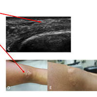
Ultrasound imaging of a scar on the knee: Sonopalpation for fascia and subcutaneous tissues
Submitted: 21 February 2020
Accepted: 29 February 2020
Published: 1 April 2020
Accepted: 29 February 2020
Abstract Views: 1153
PDF: 839
HTML: 227
HTML: 227
Publisher's note
All claims expressed in this article are solely those of the authors and do not necessarily represent those of their affiliated organizations, or those of the publisher, the editors and the reviewers. Any product that may be evaluated in this article or claim that may be made by its manufacturer is not guaranteed or endorsed by the publisher.
Authors
Department of Neurosciences, Institute of Human Anatomy, University of Padova, Italy.
RUSK Rehabilitation, New York University School of Medicine, New York, New York, United States.
Department of Neurosciences, Institute of Human Anatomy, University of Padova, Italy.
Hacettepe University Medical School, Department of Physical and Rehabilitation Medicine Ankara, Turkey.
Persistent scar pain associated with healed surgical incisions after a trauma is a common and potentially debilitating type of fascial pain. At present, there is no universally effective treatment for persistent surgical or post-trauma scar pain. Herein we describe the successful objective diagnosis of debilitating scar pain by Ultrasound (US) imaging. The sonopalpation of the fasciae and subcutaneous tissues seems to be relevant to diagnose the real cause of the pain and why not to monitor the treatment.
Franchignoni F, Giordano A, Vercelli S, Bravini E, Stissi V, Ferriero G. Rasch Analysis of the Patient and Observer Scar Assessment Scale in Linear Scars: Suggestions for a Patient and Observer Scar Assessment Scale v2.1. Plast Reconstr Surg. 2019 Dec;144(6):1073e-1079e.
DOI: https://doi.org/10.1097/PRS.0000000000006265
Vercelli S, Ferriero G, Sartorio F, Cisari C, Bravini E. Clinimetric properties and clinical utility in rehabilitation of postsurgical scar rating scales: A systematic review. Int J Rehabil Res. 2015;38:279–286.
DOI: https://doi.org/10.1097/MRR.0000000000000134
Van der Wal MB, Verhaegen PD, Middelkoop E, van Zuijlen PP. A clinimetric overview of scar assessment scales. J Burn Care Res. 2012;33:e79–e87.
DOI: https://doi.org/10.1097/BCR.0b013e318239f5dd
Pirri C, Stecco C, Fede C, Macchi V, Özçakar L. Ultrasound Imaging of the Fascial Layers: You See (Only) What You Know. J Ultrasound Med. 2019 Oct 23. doi: 10.1002/jum.15148. [Epub ahead of print]
DOI: https://doi.org/10.1002/jum.15148
Özçakar L, Kara M, Chang KV, Çarl AB, Akkaya N, Tok F, Chen WS, Wang TG, Tekin L, Ulaşl AM, Chen CP, Çapkn E, De Muynck M. Nineteen reasons why physiatrists should do musculoskeletal ultrasound: EURO-MUSCULUS/USPRM recommendations. Am J Phys Med Rehabil. 2015 Jun;94(6):e45-9. doi: 10.1097/PHM.0000000000000223.
DOI: https://doi.org/10.1097/PHM.0000000000000223
Silverberg R, Johnson J, Moffat M. The effects of soft tissue mobilization on the immature burn scar: Results of a pilot study. J Burn Care Rehabil. 1996;17:252–259.
DOI: https://doi.org/10.1097/00004630-199605000-00013
Özçakar L, Kara M, Chang KV, et al. EURO-MUSCULUS /USPRM basic scanning protocols for knee. Eur J Phys Rehabil Med 2015;51:641-6.
Mustoe TA, Cooter RD, Gold MH, Hobbs FD, Ramelet AA, Shakespeare PG, Stella M, Te´ot L, Wood FM, Ziegler UE. International clinical recommendations on scar management. Plast Reconstr Surg 2002;110:560–571.
DOI: https://doi.org/10.1097/00006534-200208000-00031
Vercelli S1, Ferriero G, Sartorio F, Stissi V, Franchignoni F. How to assess postsurgical scars: a review of outcome measures. Disabil Rehabil. 2009;31(25):2055-63.
DOI: https://doi.org/10.3109/09638280902874196a
Roques C, Te´ot L. A critical analysis of measurements used to assess and manage scars. Int J Low Extrem Wounds 2007;6:249–253.
DOI: https://doi.org/10.1177/1534734607308249
Atiyeh BS. Nonsurgical management of hypertrophic scars: evidence-based therapies, standard practices, and emerging methods. Aesth Plast Surg 2007;31:468–492.
DOI: https://doi.org/10.1007/s00266-006-0253-y
Te´ot L. Scar evaluation and management: recommendations. Eur Tissue Repair Soc Bull 2005;12:1–11.
Truong PT, Lee JC, Soer B, Gaul CA, Olivotto IA. Reliability and validity testing of the Patient and Observer Scar Assessment Scale in evaluating linear scars after breast cancer surgery. Plast Reconstr Surg 2007;119:487–494.
DOI: https://doi.org/10.1097/01.prs.0000252949.77525.bc
Powers PS, Sarkar S, Goldgof DB, Cruse CW, Tsap LV. Scar assessment: current problems and future solutions. J Burn Care Rehabil 1999;20:54–60.
DOI: https://doi.org/10.1097/00004630-199901001-00011
Curtin C, Carrol I. Cutaneous neuroma physiology and its relationship to chronic pain. J Hand Surg (USA) 2009; 34:1334-1336.
DOI: https://doi.org/10.1016/j.jhsa.2009.04.003
Lancerotto L., Stecco C., Macchi V., Porzionato A., Stecco A., De Caro R. Layers of the abdominalwall: anatomical investigation of subcutaneous tissue and superficial fascia. Surg. Radiol. Anat. 2011.33 (10), 835–842.
Stecco C. 2015. Functional Atlas of the Human Fascial System. UK:Elsevier Health Sciences.
Stecco C, Pirri C, Fede C, Fan C, Giordani F, Stecco L, Foti C, De Caro R. Dermatome and fasciatome. Clin Anat. 2019 May 14. doi: 10.1002/ca.23408. Barford A, Dorling D, Smith G, Shaw M. Life expectancy: women now on top everywhere. BMJ., 2006;332(7545):808.
How to Cite
Pirri, C., Stecco, A., Fede, C., De Caro, R., Stecco, C., & Özçakar, L. (2020). Ultrasound imaging of a scar on the knee: Sonopalpation for fascia and subcutaneous tissues. European Journal of Translational Myology, 30(1), 150–153. https://doi.org/10.4081/ejtm.2019.8909
PAGEPress has chosen to apply the Creative Commons Attribution NonCommercial 4.0 International License (CC BY-NC 4.0) to all manuscripts to be published.