Quantitative evaluation of CT scan images to determinate the prognosis of COVID-19 patient using deep learning

Accepted: 13 July 2023
HTML: 2
All claims expressed in this article are solely those of the authors and do not necessarily represent those of their affiliated organizations, or those of the publisher, the editors and the reviewers. Any product that may be evaluated in this article or claim that may be made by its manufacturer is not guaranteed or endorsed by the publisher.
Authors
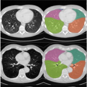
The purpose of this research is to evaluate the accuracy of AI-assisted quantification in comparison to conventional CT parameters reviewed by a radiologist in predicting the severity, progression, and clinical outcome of disease. The current study is a cross-sectional study that was conducted on patients with the diagnosis of COVID-19 and underwent a pulmonary CT scan between August 23th, 2021 to December 21th, 2022. The initial CT scan on admission was used for imaging analysis. The presence of ground glass opacity (GGO), and consolidation were visually evaluated. CT severity score was calculated according to a semi-quantitative method. In addition, AI based quantification of GGO and consolidation volume were also performed. 291 patients (mean age: 64.7 ± 7; 129 males) were included. GGO + consolidation was more frequently revealed in progress-to-severe group whereas pure GGO was more likely to be found in non-severe group. Compared to non-severe group, patients in progress-to-severe group had larger GGO volume percentage (40.6%± 11.9%versus 21.7%± 8.8%, p ˂0.001) as well as consolidation volume percentage (4.8% ± 2% versus 1.9% ± 1%, p < 0.001). Among imaging parameters, consolidation volume percentage and the largest area under curve (AUC) in discriminating non-severe from progress-to-severe group (AUC = 0.91, p < 0.001). According to multivariate regression, consolidation volume was the strongest predictor for disease progression. In conclusion, the consolidation volume measured on the initial chest CT was the most accurate predictor of disease progression, and a larger consolidation volume was associated with a poor clinical outcome. In patients with COVID-19, AI-assisted lesion quantification was useful for risk stratification and prognosis evaluation.
How to Cite

This work is licensed under a Creative Commons Attribution-NonCommercial 4.0 International License.
PAGEPress has chosen to apply the Creative Commons Attribution NonCommercial 4.0 International License (CC BY-NC 4.0) to all manuscripts to be published.

 https://doi.org/10.4081/ejtm.2023.11571


