Reviews
Vol. 25 No. 1 (2015): News on Muscle Imaging
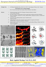
Skeletal muscle triad junction ultrastructure by Focused-Ion-Beam milling of muscle and Cryo-Electron Tomography

Publisher's note
All claims expressed in this article are solely those of the authors and do not necessarily represent those of their affiliated organizations, or those of the publisher, the editors and the reviewers. Any product that may be evaluated in this article or claim that may be made by its manufacturer is not guaranteed or endorsed by the publisher.
All claims expressed in this article are solely those of the authors and do not necessarily represent those of their affiliated organizations, or those of the publisher, the editors and the reviewers. Any product that may be evaluated in this article or claim that may be made by its manufacturer is not guaranteed or endorsed by the publisher.
Received: 6 November 2014
Published: 15 January 2015
Published: 15 January 2015
3405
Views
1440
Downloads
Downloads
Download data is not yet available.
Supporting Agencies
National Institutes of Health, New York State Department of HealthHow to Cite
1.
Wagenknecht T, Hsieh C, Marko M. Skeletal muscle triad junction ultrastructure by Focused-Ion-Beam milling of muscle and Cryo-Electron Tomography. Eur J Transl Myol [Internet]. 2015 Jan. 15 [cited 2026 May 11];25(1):49-56. Available from: https://www.pagepressjournals.org/bam/article/view/bam.2015.1.49
PAGEPress has chosen to apply the Creative Commons Attribution NonCommercial 4.0 International License (CC BY-NC 4.0) to all manuscripts to be published.